

Breech presentation Treatment in Nagpur: The magnificent journey of human gestation represents an intricate, beautifully orchestrated biological choreography uniting maternal physiology and fetal development over forty weeks of profound transformation. As expectant parents approach the culmination of pregnancy, anticipation builds naturally around the moment of birth and the physical posture the baby assumes within the intrauterine sanctuary. While the vast majority of fetuses spontaneously rotate into a head-down (cephalic) orientation by the third trimester, a small percentage of babies settle into a breech presentation—positioned feet-first or buttocks-first rather than head-first. In the vibrant, medically progressive city of Nagpur, elite maternal-fetal healthcare infrastructure provides expectant mothers facing breech presentations with world-class diagnostic evaluation, advanced turning techniques, and specialized delivery planning. Whether you have managed your gestational journey under comprehensive Pregnancy Care protocols, navigated specialized surveillance for a High Risk Pregnancy in Nagpur, addressed early-trimester spotting requiring Vaginal bleeding Treatment, evaluated anatomical development via an Anomaly Scan in Nagpur or third-trimester Growth Scan in Nagpur, monitored placental hemodynamics through Obstetric Doppler in Nagpur, sought genetic clarity via Amniocentesis in Nagpur, prepared for complex delivery within an elite High Risk Delivery with ICU backup in Nagpur suite, or established robust preventive baselines during preconception counseling like Pre Conception Counselling, understanding the true clinical scope of breech presentation management is essential for absolute peace of mind. Cultural anxiety, generalized misconceptions regarding breech deliveries, or unfamiliarity with modern obstetric turning techniques can sometimes cause unnecessary worry. It is vital to recognize that modern advanced obstetrics prioritizes proactive ultrasound surveillance, safe external cephalic version protocols, and individualized delivery planning to ensure optimal safety without relying on chemical or pharmaceutical drug interventions. Finding an expert, empathetic Gynecologist in Nagpur is the foundational step in receiving precise clinical counseling, elite hospital coordination, and personalized guidance tailored to your unique anatomical and physiological profile. To truly comprehend the profound scope of breech presentation treatment, one must explore the fundamental biomechanics of fetal positioning, anatomical variations of breech posture, diagnostic imaging modalities, external cephalic version maneuvers, planned surgical versus vaginal delivery pathways, postpartum recovery, and holistic urogenital restoration. Consequently, approaching breech presentation requires a multi-dimensional, deeply supportive framework that honors clinical excellence, uncompromising safety, and holistic maternal-fetal well-being.
The Physiology of Fetal Positioning and Gravitational Alignment
To effectively approach breech presentation treatment, it is medically necessary to explore the natural physiological mechanisms governing fetal movement and gravitational alignment throughout gestation. During the first and second trimesters, the fetus is relatively small compared to the generous volume of surrounding amniotic fluid, allowing it to move freely, tumble, and shift positions frequently within the uterine cavity.
As pregnancy progresses into the third trimester, the physics of the intrauterine environment change dramatically. The fetus grows larger and heavier, while the relative volume of amniotic fluid decreases proportionally. Due to the pear-shaped anatomy of the human uterus—where the wider fundus accommodates the heavier fetal breech and lower extremities, while the narrower lower segment cradles the denser fetal head—gravitational forces naturally encourage the baby to perform a somersault, settling into a stable cephalic presentation by the thirty-sixth week. When this spontaneous rotation does not occur, the baby remains in a breech posture. Understanding this physiological baseline empowers clinical teams to assess positioning accurately and intervene with appropriate, timely strategies.
Defining Breech Presentation and Its Anatomical Variations
A breech presentation occurs when the fetus occupies a longitudinal lie within the uterus with the buttocks or lower extremities positioned closest to the maternal cervical os. Depending on the exact flexion of the fetal hips and knees, obstetricians classify breech presentations into three distinct anatomical variations:
- Frank Breech (Most Common): The fetal hips are flexed with both legs extended straight upward toward the chest, with the feet resting near the baby’s face. This posture resembles a V-shape and accounts for the majority of breech presentations, offering a compact anatomical profile.
- Complete Breech: Both the fetal hips and knees are fully flexed, with the baby sitting cross-legged inside the uterine cavity (similar to a tailor-sitting posture), positioning the buttocks and feet downward toward the cervix.
- Footling or Incomplete Breech: One or both feet are positioned below the buttocks, extending downward into the lower uterine segment. This represents the least common and most complex breech variation, requiring careful clinical evaluation due to the potential risk of umbilical cord prolapse.
Recognizing these specific anatomical variations allows the maternal-fetal medicine specialist to tailor clinical management precisely to the physical characteristics of the gestation.
Antenatal Diagnosis and Advanced Imaging Protocols
Accurately diagnosing a breech presentation is a cornerstone of modern prenatal care, enabling obstetricians to plan management strategies well before the onset of labor. While routine physical examination using Leopold maneuvers (palpating the maternal abdomen) can suggest fetal orientation, definitive confirmation relies upon advanced ultrasonography.
The core diagnostic imaging protocols include:
- Third-Trimester Biometric Scans: Utilizing a detailed Growth Scan in Nagpur to confirm fetal presentation, assess amniotic fluid volume, estimate fetal weight, and evaluate placental location.
- Detailed Anatomical Correlation: Ensuring that findings are cross-referenced with previous imaging such as the mid-trimester Anomaly Scan in Nagpur to rule out any structural variations.
- Hemodynamic and Vascular Assessment: Performing Obstetric Doppler in Nagpur studies to confirm optimal umbilical and cerebral blood flow, ensuring the fetus is thriving despite its non-cephalic posture.
This rigorous diagnostic suite ensures that every clinical decision is grounded in precise, real-time anatomical data.
Etiologies and Associated Risk Factors
While many breech presentations occur spontaneously in structurally normal pregnancies without an identifiable cause, certain physiological and anatomical factors can influence fetal positioning.
The primary factors associated with breech presentation include:
- Amniotic Fluid Volume Variations: Conditions involving excessive fluid (polyhydramnios) permit excessive fetal mobility, while diminished fluid (oligohydramnios) restricts the baby’s ability to turn into a cephalic posture.
- Uterine Anatomical Abnormalities: Structural features such as uterine fibroids, septate uteri, or prior scar tissue can alter the internal contour of the cavity. For women with prior histories evaluated during Uterine polyps Treatment or endocrine variations managed under structured PCOS Treatment protocols, careful structural monitoring is paramount.
- Placental Location: A low-lying placenta or placenta previa can occupy the lower uterine space, preventing the fetal head from engaging downward.
- Multiple Gestations: Carrying twins or higher-order multiples naturally limits intrauterine space, frequently resulting in one or both fetuses adopting a breech orientation.
- Fetal Maturity and Gestational Age: Pregnancies evaluated prior to thirty-four weeks naturally exhibit a higher incidence of breech positioning that resolves spontaneously as term approaches.
Identifying these underlying contributors allows the clinical team to approach breech management with comprehensive foresight.
External Cephalic Version (ECV): The Gentle Turning Technique
For mothers carrying a breech baby near term (typically between thirty-six and thirty-seven weeks), External Cephalic Version (ECV) offers an exceptionally effective, non-surgical method to encourage the baby to turn into a head-down cephalic presentation.
The ECV Procedure Protocol: Performed in a fully equipped obstetric facility, ECV involves the skilled application of gentle, firm manual pressure on the maternal abdominal wall by an experienced obstetrician. Guided by real-time ultrasound, the clinician guides the fetus through a forward or backward somersault, moving the breech out of the pelvis and rotating the head downward.
Safety and Monitoring Standards: Throughout the procedure, fetal heart rate and maternal vital signs are continuously monitored via cardiotocography. If any signs of transient fetal distress occur, the maneuver is immediately paused, allowing the fetus to return to its original position. Success rates for ECV vary based on parity, amniotic fluid volume, and placental placement, offering many women the opportunity to achieve a safe vaginal delivery.
Evaluating Delivery Pathways: Planned Cesarean Section Versus Vaginal Breech Delivery
When a breech presentation persists despite turning efforts or clinical preference, determining the safest mode of delivery is a critical decision made in close collaboration between the obstetrician and the expectant parents.
Planned Cesarean Delivery: Backed by extensive clinical data demonstrating superior safety profiles for breech infants, a scheduled elective cesarean section performed around thirty-nine weeks is frequently recommended as the primary mode of delivery. This approach eliminates the mechanical risks associated with vaginal breech delivery, ensuring a controlled birth environment with integrated High Risk Delivery with ICU backup in Nagpur readiness.
Strict Criteria for Managed Vaginal Breech Delivery: In select cases—such as a frank breech presentation, proven adequate maternal pelvic dimensions, an estimated fetal weight within safe parameters, and continuous intrapartum monitoring—a carefully managed vaginal breech delivery may be considered under the direct supervision of an expert obstetric team skilled in breech maneuvers.
Postpartum Recovery, Pelvic Floor Preservation, and Urogenital Health
Whether a breech presentation culminates in a planned cesarean section or a managed vaginal birth, dedicating intentional care to postpartum recovery is essential for supporting maternal tissue restoration.
Postpartum Healing and Rest: Following delivery, the maternal body undergoes rapid involution. Ensuring adequate physical rest, proper wound care, and consistent hydration supports rapid recovery and physiological normalization.
Preventing Long-Term Urogenital Complications: The physical strain of advanced gestation and delivery places significant stress on the pelvic floor muscles and supportive fascial ligaments. Ensuring proper postpartum healing prevents long-term urogenital complications, protecting against future issues such as postpartum stress urinary leakage requiring Stress incontinence Treatment or fascial descents managed under Uterine prolapse Treatment protocols. Integrating gentle pelvic floor rehabilitation into postpartum recovery restores core strength and long-term pelvic harmony.
Nutritional Excellence, Whole Foods, and Restorative Wellness
Nourishing the maternal body through the final weeks of gestation and supporting postpartum tissue regeneration requires an uncompromising commitment to wholesome nutrition and natural dietary excellence.
Nutrient-Dense Whole Foods and Botanical Support: Emphasizing an abundant variety of deeply pigmented antioxidant vegetables, dark leafy greens, nutrient-dense whole grains, legumes, seeds, and fresh organic fruits provides the vital micronutrients, vitamins, and minerals required for rapid cellular repair, tissue regeneration, and metabolic vitality.
Hydration and Metabolic Restoration: Maintaining consistent daily hydration with pure water, fresh coconut water, and mineral-rich herbal infusions supports expanded blood volume normalization and efficient metabolic waste elimination, ensuring smooth circulatory function across all organ systems.
Emotional Sanctuary and Stress Reduction: Honoring the body’s need for deep psychological and physical rest regulates maternal cortisol levels, fostering a peaceful internal environment that accelerates healing and strengthens the sacred bond between mother and newborn.
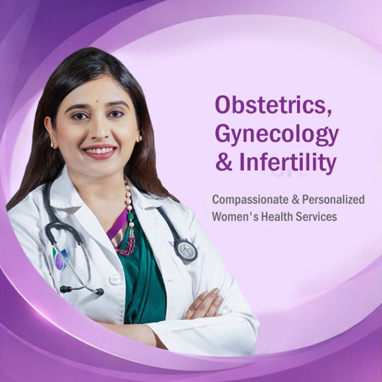
Meet Your Expert : Dr. Samiksha Jain
Dr. Samiksha Jain
Obstetrician, Gynecologist & Fertility Specialist
MBBS, MD (Obs/Gyn) Gold Medalist
Fellowship in Reproductive Medicine (Bangalore),
Fellowship in Minimal Access Surgery (Bangalore),
Advanced Diploma in Reproductive Medicine (Germany)
Dr. Samiksha Jain is a highly accomplished Obstetrician, Gynecologist, and Fertility Specialist known for her compassionate approach and excellence in women’s healthcare. A Gold Medalist in MD (Obstetrics & Gynecology), she combines academic brilliance with advanced clinical expertise to provide the highest standard of care to her patients. Dr. Samiksha Jain is an esteemed Gynecologist dedicated to empowering women through natural, comprehensive, and deeply personalized health protocols. With her profound expertise in high-risk obstetrics, maternal-fetal medicine, and breech presentation management, she offers state-of-the-art clinical support, external cephalic version coordination, and expert delivery planning, ensuring a safe, deeply reassuring, and beautifully joyful gestation and childbirth journey for every mother.
Frequently Asked Questions (FAQs)
1. What is a breech presentation, and why do some babies remain in this position?
A breech presentation occurs when a baby is positioned feet-first or buttocks-first rather than head-down near term. This can be influenced by amniotic fluid volume, uterine anatomy, or placental placement.
2. What are the primary anatomical variations of breech presentation?
The three main variations are frank breech (legs extended upward), complete breech (hips and knees flexed), and footling breech (feet positioned downward).
3. How is a breech presentation diagnosed during pregnancy?
Diagnosis is confirmed through third-trimester ultrasound imaging, such as a specialized Growth Scan in Nagpur, which evaluates fetal orientation and amniotic fluid volume.
4. What is External Cephalic Version (ECV), and when is it performed?
ECV is a manual turning procedure performed under ultrasound guidance around thirty-six to thirty-seven weeks of gestation to encourage a breech baby to rotate into a head-down position.
5. Is a cesarean section always required for a breech presentation?
While a scheduled elective cesarean section is frequently recommended for safety, select cases meeting strict clinical criteria may be evaluated for a managed vaginal breech delivery.
6. How does pre-conception planning support healthy gestational outcomes?
Engaging in proactive Pre Conception Counselling establishes a pristine physiological baseline, optimizing maternal health and metabolic preparedness before pregnancy begins.
7. What nutritional steps support recovery after delivery?
Consuming nutrient-dense whole foods rich in vitamins, minerals, and antioxidants, alongside consistent hydration, supports rapid tissue repair and overall physical restoration.
8. When should I book my initial prenatal and delivery consultations in Nagpur?
You should schedule your consultations early in your pregnancy, allowing your obstetrician to integrate elite hospital planning and breech management coordination seamlessly into your comprehensive Pregnancy Care roadmap.
Disclaimer: The comprehensive information provided in this extensive article is strictly for educational, informational, and awareness purposes only. It is not intended to be a substitute for professional medical advice, clinical diagnosis, or specialized therapeutic treatment. Always seek the direct, personalized advice of your physician or qualified healthcare provider regarding any medical condition, anatomical concerns, or prior to initiating any new dietary change, physical therapy, or surgical procedure.